It looks like you're using an Ad Blocker.
Please white-list or disable AboveTopSecret.com in your ad-blocking tool.
Thank you.
Some features of ATS will be disabled while you continue to use an ad-blocker.
1
share:
Although pictures of the brain usually gross me out, these were actually refreshing and quite interesting as well. One or two of the pictures was
taken with the help of an ingenious idea, whose inventors recieved the 2008 Nobel Prize in chemistry. If you're interested in reading more about this
invention than whats written in the caption of the pictures, I'm providing a link at the bottom for you.


Source: More Pictures Here: The Brain Is Ready for Its Close-up
Nobel Prize in Chemistry 2008 - Press Release

Through the 20th century, neuroscientists gradually tracked particular brain functions to specific regions. Sadly, the most efficient way to determine such a linkage was to find a patient with an injury to a specific region of his brain, and then to establish which of the patient's faculties were impaired.
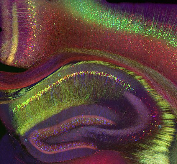
One of these patients was H.M., an epileptic who went under the surgeon's knife at the age of 27--the doctors stopped his seizures, but they also cut into a brain region called the hippocampus and inadvertently destroyed his ability to form new memories. For the rest of his life, until he died in 2008 at the age of 82, he could only hold a thought for about 20 seconds. Yet H.M. could clearly remember things from before his surgery, and could also form new "motor memories"--for example, after many repetitions he performed a difficult drawing task with ease, but had no memory of doing it before. Such results helped researchers understand the role of the hippocampus, which is shown in cross-section at the bottom half of this 2005 image.

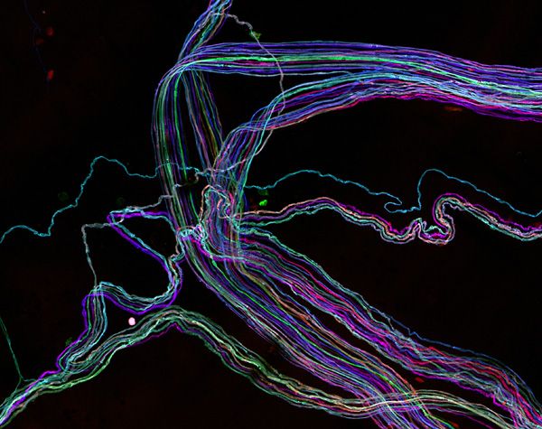
We have luminescent jellyfish to thank for one advance in neuroscience. By sticking the genetic code for the jellyfish's green fluorescent protein in cells, researchers made them glow green (a neat trick that won the 2008 Nobel Prize in chemistry). Most importantly, this could be done in living animals and tissues, allowing researchers to watch neurons in action. To sort out the tangle of brain cells, molecular chemists next searched out fluorescent proteins in other colors, and found innovative ways to make neurons glow in slightly different hues.
This "brainbow" image traces the long axons of individual neurons in a gorgeous array of colors. The image, produced in 2007, shows motor neuron axons stretching away to the muscles they control.
Source: More Pictures Here: The Brain Is Ready for Its Close-up
Nobel Prize in Chemistry 2008 - Press Release
reply to post by Droogie
Well I never thought I'd see intimate and brain in the same sentence but it is fitting here. These are awesome shots and the "brainbow" sounds crazy, with jellyfish injections, but it sure is purdy.
nice find!
Peace,
spec
Well I never thought I'd see intimate and brain in the same sentence but it is fitting here. These are awesome shots and the "brainbow" sounds crazy, with jellyfish injections, but it sure is purdy.
nice find!
Peace,
spec
reply to post by speculativeoptimist
Yeah, it's quite fascinating, and those really are some unique pictures. Fascinating how they came up with that method of looking at an organ.
Yeah, it's quite fascinating, and those really are some unique pictures. Fascinating how they came up with that method of looking at an organ.
new topics
-
An Interesting Conversation with ChatGPT
Science & Technology: 13 minutes ago -
World's Best Christmas Lights!
General Chit Chat: 10 hours ago -
Drone Shooting Arrest - Walmart Involved
Mainstream News: 10 hours ago -
Can someone 'splain me like I'm 5. Blockchain?
Science & Technology: 11 hours ago
top topics
-
Georgia appeals court disqualifies DA Fani Willis from Trump election interference case
US Political Madness: 15 hours ago, 25 flags -
Have you noticed?? Post Election news coverage...
World War Three: 13 hours ago, 12 flags -
Squirrels becoming predators
Fragile Earth: 12 hours ago, 10 flags -
Drone Shooting Arrest - Walmart Involved
Mainstream News: 10 hours ago, 10 flags -
Can someone 'splain me like I'm 5. Blockchain?
Science & Technology: 11 hours ago, 7 flags -
World's Best Christmas Lights!
General Chit Chat: 10 hours ago, 7 flags -
Labour's Anti-Corruption Minister Named in Bangladesh Corruption Court Papers
Regional Politics: 12 hours ago, 6 flags -
An Interesting Conversation with ChatGPT
Science & Technology: 13 minutes ago, 0 flags
1
