It looks like you're using an Ad Blocker.
Please white-list or disable AboveTopSecret.com in your ad-blocking tool.
Thank you.
Some features of ATS will be disabled while you continue to use an ad-blocker.
share:
reply to post by mikemck1976
Of course you are correct. Greer just makes money off rehashing the same stuff every year at the press conference. Good post. Thanks
Of course you are correct. Greer just makes money off rehashing the same stuff every year at the press conference. Good post. Thanks
Originally posted by mikemck1976
Mr. Ripley found the shrunken body in Peru and named it “Atta-Boy”, after the nearby Atacama Desert, the same desert where Dr. Steven Greer would eventually find the humanoid body in Sirius.
Here's a video of Lee Speigel from the Huffington Post talking about it.
link
Hi smal-entity fans.
Come on !!!
Don't you see that the shadows do not match !?
In the first photo, it is impossible to have the shadow of Ripley's head
on the wall on the right side of his mouth !!
And in the photo of the big hand, check carefuly for the shadows. . .
they are not consistant, the-little-being related to the hand and the wall behind !!
Blue skies.
reply to post by seabhac-rua
Looks fake to me. Where are the DNA samples proving it is real? How about some X-rays of the miniature person Mr. Ripley is holding? Even then, that doesn't explain why the 'alien' has 9 ribs instead of 12 (on each side). Face it, the two are still not at all relatable. There are just too many discrepancies between the two.
Looks fake to me. Where are the DNA samples proving it is real? How about some X-rays of the miniature person Mr. Ripley is holding? Even then, that doesn't explain why the 'alien' has 9 ribs instead of 12 (on each side). Face it, the two are still not at all relatable. There are just too many discrepancies between the two.
Originally posted by Bilk22
reply to post by cavtrooper7
How was my post off topic? I asked the OP if he knew how shrunken heads were made. He clearly doesn't and should read more on the subject before starting a thread that was obviously calling on the veracity of not only Greer, but the scientists at Stanford. IMO the OP needed to be called out and I didn't do it in any ill manner.
Hm, I see no appology from the mods no wonder ATS is going downhill with yet another example of uneccassary censorship.
Absolute power corrupts absolutely
reply to post by mikemck1976
Very nice find! S & F
(can you provide a link to the Ripley's Atta-Boy)
Very nice find! S & F
(can you provide a link to the Ripley's Atta-Boy)
reply to post by Staroth
Here you go
Believe it or Not: The Atacama “Alien” is Human, and Ripley Knew It First
(can you provide a link to the Ripley's Atta-Boy)
Here you go
Believe it or Not: The Atacama “Alien” is Human, and Ripley Knew It First
Why can't you shrink bones?
It might sound like a pretty stupid stupid question.. but then again is there really such a thing as a stupid question?
"The exact mechanisms by which bone development is triggered remains unclear, but it involves growth factors and cytokines in some way."
en.wikipedia.org...
Apparently people are already working on "reverse ossification" which can INCREASE height and growth of bones through microfracture surgery, so shrinking bones doesn't sound all that far fetched to me.
browse.feedreader.com...
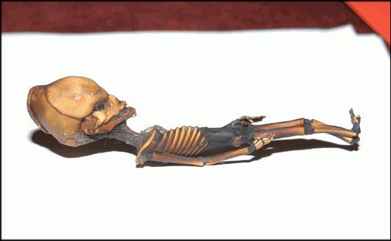
is it just me or does the picture of the "skeleton" look its been drawn, or is a drawing?
on the other hand, think about how much more room we would have if this was done prior to burial.
i want one.
on the other hand, think about how much more room we would have if this was done prior to burial.
i want one.
look closely at Greer's find there are no knee caps or elbow caps. this tells us it is a newborn baby. newborns bones are not solidified for a few
days and are very supple and can be shrunk they are more like tissue for the first 24 hours. You will also notice that the Sternum is missing another
sign it is a newborn baby as the sternum takes a while to connect the ribs and grow if it were there at birth it would hurt the mother greatly and
kill the child during natural birth.
I remember doing a little reading on shrunken heads a long time ago, and if I remember correctly, they actually remove the skull altogether, because
the process of shrinking does not work on something as hard and dense as bone. Therefore it is virtually impossible for your idea to be feasible. I
would not doubt that Ripley's specimen is just as fake as Greer's. I know that Greer likely commands more respect that others within the same
community, but I just find it hard to believe that this skeleton is actually authentic. If it is, why hasn't it gotten public recognition? This is
something that the media would NOT want to hide, and they would be all over it, IF it could be proven to be real. At least that is my opinion.
reply to post by mikemck1976
interesting, but if you watched the documentary you would know that doctors from stanford noted many more oddities than simply size. number of ribs, the massive cranium, etc... I feel like if it was 100% human (if this thing is real in the first place), doctors could figure it out.
The dessert coincident is notable though.
reply to post by mikemck1976
Shrunken head technique involves removing the bones -- in the case of a head, the skull. The "Sirius/Greer" body appears to include the skeleton, so I don't think it is a shrunken body -- not even the shrunken body of an initially small human.
Shrunken head technique involves removing the bones -- in the case of a head, the skull. The "Sirius/Greer" body appears to include the skeleton, so I don't think it is a shrunken body -- not even the shrunken body of an initially small human.
Originally posted by mikemck1976
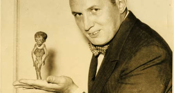
Pick up and flip though Ripley's Search for the Shrunken Heads: and Other Curiosities and you will find the image above of Ripley holding a familiar humanoid. According to his notes, the oddity turned out to be a person who was chosen for a full-body “reduction” rather than just a shrinking of the head.
Where did Mr. Ripley find this little guy?
Mr. Ripley found the shrunken body in Peru and named it “Atta-Boy”, after the nearby Atacama Desert, the same desert where Dr. Steven Greer would eventually find the humanoid body in Sirius.
Dr. Steven Greer's Sirius Alien
I think that the two "humanoids" are very similar and considering that both came from the same area, leads me to believe that Dr. Steven Greer's Sirius Alien is just another example of Robert Ripley's “Atta-Boy”...
Some of us still read books Mr. Greer!
edit on 4-6-2013 by mikemck1976 because: (no reason given)
Very interesting find although there are still some inconsistencies with such an observation as the humanoid that Greer has sent to Stanford university for DNA sampling has had some obscure results. It matches 91% with our DNA but as you know chimps are 98% alike in their DNA structuring and we can see the vast differences between them and us. Of course Greer is a bit of a sensationalist but when it comes down to the hard science this thing is a mystery to professionals regarded as some of the top in their field. Now whether it is the product of shrinking and the method of such an action that could manipulate the DNA properties of such a human is another venture that could be explored. I suppose I am a bit bias in that I want that little bugger to an alien so that humanity can realize just how small we really are in the cosmos, you know, bring some humility so our egos may stop figuratively strangling each other. The probability of other life out there is 100% if you ask me, the question we should be asking is if there are beings out there that have mastered physics to visit us.
Atacama mystery
reply to post by gortex
While one would assume that bone density would increase with mummification, a recent study proved that to not be the case: Mummified Cat
And the conclusion...
My money is still on the Stanford team...
peace,
AB
While one would assume that bone density would increase with mummification, a recent study proved that to not be the case: Mummified Cat
Nelson, associate dean of research and operations in Western’s Department of Anthropology, and associate dean in the Faculty of Social Science, has led the Yes investigation, looking to determine whether changes that happen to tissues are part of the pathological process or related to mummification. In other words, is the density of the vertebrae, observed in radiographs of Ramses II, indicative of him having suffered from AS? Or, is the density a result of the mummification process? “We’re looking at the osteobiography of a mummy. We’re trying to tell the story of that person’s life through the analysis of bones and tissues; we want to get as accurate a picture of their life as we can, that we can properly diagnose the disease process and properly differentiate from (the mummification process),” Nelson explained... The goal was to see what changes can be observed in tissues and how long it takes for such changes to occur. Once mummification was complete, researchers examined Yes with MR (magnetic resonance) scans and clinical CT (computerized tomography) imaging, in order to see beneath the wrappings and observe changes to tissues over time.
And the conclusion...
The results of the scans showed a rapid shrinking and a decrease in tissue density, Nelson said, noting the expectation was that tissues would increase in density, not get softer. What this means, Nelson said, is that if we observe increased density in tissue of a mummy, researchers can be confident that it represents real physiological issues, ones not part of the mummification process. “If we see something that is markedly more dense in a mummy, we can be sure it is pathology,” he said.
My money is still on the Stanford team...
peace,
AB
edit on 6-6-2013 by AboveBoard because: (no reason given)
reply to post by AboveBoard
Hi AB
Interesting article but as no time scale is given for the test it's hard to be able to draw any conclusions , the test subject and Ata would also of undergone different processes as Ata was buried in the desert for an undetermined length of time but we don't know if the test subject was buried at all , it also appears well wrapped but we don't know if or how Ata was wrapped .
I think we'll have to sit in our respective camps and wait for more tests until we can finally put this one to bed although I still believe the balance of probability is that Ata is an early stage foetus
edit on 6-6-2013 by gortex because: (no reason given)
Further research on mummification of fetal remains and the process of determining age relevant to Ata:
FETAL MUMMY STUDY
Has bone growth been used before in determining the age of fetal mummies? Yes!
This radiological / CT study of two mummified fetus remains clearly states that ossification in the knees is used as part of the factors in determining age.
Mummified Daughters of King Tutankhamun: Archeologic and CT Studies: American Journal of Roentgenology: Vol. 197, No. 5
NOTE: "secondary ossification centers" refers to the place where future bone growth would take place NOT actual epiphyseal plate growth - Close to the time of birth (around 36-42 weeks) these secondary ossification centers appear. Endochoral ossification & fetal growth
BONE GROWTH TIMETABLE & PROCESS:
The process and timetable for ossification: Ossification - Human Timetable Ossification is when the body builds new bone - the knee is one of the "secondary centers" and does not go through the process of ossification until after birth.
The specific form of ossification related to the knees and long bones is "endochondral ossification." There are two centers for this, the first being "Primary" growth which begins in embryonic development, and the "secondary" which is where bone growth occurs after birth and at a certain rate of formation that are called "epiphyseal standards." These are the standards used to determine "Ata's" age, by Dr. Ralph Lachman from Stanford.
The epiphysis is the round end of a bone: Epiphysis
The epiphyseal plate is the "growth plate" which is found in children and adolescents: Epiphyseal Plate
Here is a site that has an awesome explanation of bone growth at the epiphyseal plate, including a slide presentation: Bone Growth
A summary -
the epiphyseal plate, or growth plate, is above the epiphysis (round end of the bone) so that joint movement can continue smoothly as growth occurs.
1) The first stage of bone growth on the epiphyseal plate is the production of hyaline cartilage which creates a space between the epiphysis and the primary length of the bone.
2) At the area the cartilage attaches to the primary length of the bone, it begins to calcify/ossify into hardened bone.
Why go through all of this? To determine for myself that 1) IF Ata was merely a fetus, then he would not have growth at the epiphyseal plates of his knees - the "secondary center" for the growth would be present, but not the actual, measurable bone growth, and 2) forensic anthropologists have used secondary ossification centers to determine the age of high-profie mummified fetal remains, and they did not see secondary growth postmortem, but only the formation of secondary centers where future growth would occur. 3) Atta is still a cool mystery.
Granted, I'm NOT an expert, but this is the best I can do right now with a half-day's research!
peace,
AB
FETAL MUMMY STUDY
Has bone growth been used before in determining the age of fetal mummies? Yes!
This radiological / CT study of two mummified fetus remains clearly states that ossification in the knees is used as part of the factors in determining age.
Mummified Daughters of King Tutankhamun: Archeologic and CT Studies: American Journal of Roentgenology: Vol. 197, No. 5
Through analysis of the reconstructed CT images of mummy 317b, we detected tooth mineralization and multiple ossifying centers around the knee, foot, sternum, and pelvis (Table 2). CT detection of secondary ossification centers around both knees of mummy 317b suggested a gestational age of approximately 38 weeks compared with known values [10]. Secondary ossification around the knees was not visualized on radiographs in a previous study of mummy 317b by Hellier and Connolly [5], who suggested a gestational age of approximately 30 weeks. More sophisticated radiographic methods of perinatal assessment of bone age with parameters such as detailed mineralization of the teeth, changes in shape in centers of ossification, and the mere presence or absence of a center have been used [11, 12]. With two of these methods, the Stempfle score and the Olsen method, we estimated gestational ages at mummification of mummy 317b of 37.3 and 39 weeks. We calculated the mean gestational age of mummy 317b to be 36.78 (SD 1.91) weeks by considering the results of the different methods we used for age estimation.
NOTE: "secondary ossification centers" refers to the place where future bone growth would take place NOT actual epiphyseal plate growth - Close to the time of birth (around 36-42 weeks) these secondary ossification centers appear. Endochoral ossification & fetal growth
BONE GROWTH TIMETABLE & PROCESS:
The process and timetable for ossification: Ossification - Human Timetable Ossification is when the body builds new bone - the knee is one of the "secondary centers" and does not go through the process of ossification until after birth.
The specific form of ossification related to the knees and long bones is "endochondral ossification." There are two centers for this, the first being "Primary" growth which begins in embryonic development, and the "secondary" which is where bone growth occurs after birth and at a certain rate of formation that are called "epiphyseal standards." These are the standards used to determine "Ata's" age, by Dr. Ralph Lachman from Stanford.
The Secondary centre
The secondary centres generally appear at the epiphysis. Secondary ossification mostly occurs after birth (except for distal femur and proximal tibia which occurs during foetal development). The epiphyseal arteries and osteogenic cells invade the epiphysis, depositing osteoblasts and osteoclasts which erode the cartilage and build bone. This occurs at both ends of long bones but only one end of digits and ribs.
The epiphysis is the round end of a bone: Epiphysis
The epiphyseal plate is the "growth plate" which is found in children and adolescents: Epiphyseal Plate
Here is a site that has an awesome explanation of bone growth at the epiphyseal plate, including a slide presentation: Bone Growth
A summary -
the epiphyseal plate, or growth plate, is above the epiphysis (round end of the bone) so that joint movement can continue smoothly as growth occurs.
1) The first stage of bone growth on the epiphyseal plate is the production of hyaline cartilage which creates a space between the epiphysis and the primary length of the bone.
2) At the area the cartilage attaches to the primary length of the bone, it begins to calcify/ossify into hardened bone.
Why go through all of this? To determine for myself that 1) IF Ata was merely a fetus, then he would not have growth at the epiphyseal plates of his knees - the "secondary center" for the growth would be present, but not the actual, measurable bone growth, and 2) forensic anthropologists have used secondary ossification centers to determine the age of high-profie mummified fetal remains, and they did not see secondary growth postmortem, but only the formation of secondary centers where future growth would occur. 3) Atta is still a cool mystery.
Granted, I'm NOT an expert, but this is the best I can do right now with a half-day's research!
peace,
AB
edit on 6-6-2013 by AboveBoard because: (no reason given)
reply to post by gortex
Hey gortex - I just spent WAY too much time researching - I just posted it (above). Let me know what you think.
It is reasonable to think that Ata is a fetus, but the more I research, the more I see how mysterious the little guy is...
Respectfully always,
AB
Hey gortex - I just spent WAY too much time researching - I just posted it (above). Let me know what you think.
It is reasonable to think that Ata is a fetus, but the more I research, the more I see how mysterious the little guy is...
Respectfully always,
AB
one word came to mind when i saw the pic of Dr. Steven Greer's Sirius Alien: Beavis
apparently I wasn't wrong:
Shocking truth exposed: The Atacama humanoid is actually Beavis

lol, j/k... \m/
apparently I wasn't wrong:
Shocking truth exposed: The Atacama humanoid is actually Beavis

lol, j/k... \m/
new topics
-
Chinese national busted in LA sending weapons to NK
World War Three: 23 minutes ago -
South Korea declares martial law for first time in 50 years over North Korea threat
Other Current Events: 2 hours ago -
Alien warfare predicted for December 3 2024
Aliens and UFOs: 6 hours ago
top topics
-
Alien warfare predicted for December 3 2024
Aliens and UFOs: 6 hours ago, 13 flags -
Statements of Intent from Incoming Trump Administration Members - 2025 to 2029.
2024 Elections: 16 hours ago, 9 flags -
Could Biden pardon every illegal alien
Social Issues and Civil Unrest: 17 hours ago, 8 flags -
South Korea declares martial law for first time in 50 years over North Korea threat
Other Current Events: 2 hours ago, 8 flags -
Stop the Presses! Turkey Soup.
Food and Cooking: 17 hours ago, 5 flags -
Chinese national busted in LA sending weapons to NK
World War Three: 23 minutes ago, 0 flags
active topics
-
Alien warfare predicted for December 3 2024
Aliens and UFOs • 30 • : WeMustCare -
Assad flees to moscow
World War Three • 48 • : cherokeetroy -
Never say Never?
Science & Technology • 47 • : Oldcarpy2 -
South Korea declares martial law for first time in 50 years over North Korea threat
Other Current Events • 19 • : onestonemonkey -
Chinese national busted in LA sending weapons to NK
World War Three • 0 • : Ravenwatcher -
Could Biden pardon every illegal alien
Social Issues and Civil Unrest • 23 • : 777Vader -
Biden pardons his son Hunter despite previous pledges not to
Mainstream News • 127 • : Flyingclaydisk -
-@TH3WH17ERABB17- -Q- ---TIME TO SHOW THE WORLD--- -Part- --44--
Dissecting Disinformation • 3465 • : 777Vader -
I thought Trump was the existential threat?
World War Three • 186 • : cherokeetroy -
Elon Musk to Make Games Great Again - XAI_GAMES Announcement Incoming.
Video Games • 24 • : ICURNVS


