It looks like you're using an Ad Blocker.
Please white-list or disable AboveTopSecret.com in your ad-blocking tool.
Thank you.
Some features of ATS will be disabled while you continue to use an ad-blocker.

More on the Pineal
page: 1share:
I know many of you here have laid down to the altar of the Pineal Gland. Out of all of the things to "worship" in a sense, it's not a bad one to
choose.
Now, mind you, we have to consider that the brain, in it's seemingly infinite complexity, is a highly differentiated device, meaning it is composed of many different macro structures, which they themselves are made up of mostly neurons and other cells which are strongly electrochemical in nature (electromagnetic).
That being said, I'd like to go into the Pineal Gland a wee bit.
Some of you are familiar with the Pineal Gland, and how it is a "third eye" in a sense. The Pineal gland has remnant features that are photo or photon recepting cells within it's body. This is due to fact that the Pineal gland is directly related to Sleep, consciousness, and the circadian cycle.
See, what happens is the Pineal Gland produces Melatonin by default. However, when the wavelengths of optical light, I believe somewhere around 520-540 nm (which is blue light) enter your eyes, those specific photons, of that specific energy, cause the Pineal Gland to shut off Melatonin production. Ever heard of blue blocker lenses? People often will wear these an hour or two before sleep so that they are sleepier.
As for dream time. Well, it's highly possible that the Pineal gland to send out specific triggers, but, if you want to get a better understanding of the matter, you need to look at what the Pineal Gland is connected to, and what those things are directly influenced by.
More on the Pineal's make up:
Wha-wha whaaaat? We're talking 4 different types of cells in this puny gland, some of which are neurons! That's one hard working gland.
Now for the Pineal's associations:
If you notice, the Pineal gland (in red) has a red "stalk" that travels down into the very center of the Cerebellum. I'm fairly certain this "Stalk" is a stalk of white matter communicating signals from the Pineal to the Arbor Vitae, which is the central white matter section of the cerebellum. The cerebellum is lightly understood to control muscle coordination, but more recent research has suggested that it sends signals to other parts of the brain, not only the body, which leads me to believe it may just be related to COORDINATION, or TIMING in general.
Furthermore, the simple fact that the pineal gland has a channel going directly into the center of the Cerebellum (which has up to 4 times more processing matter "Gray matter" than the Cerebrum, despite only making up 10% of the brain's volume), combined with the fact that the the cerebellum has, at the center, a seemingly random snake-like portion of gray matter, leads to more questions about the Pineal and it's integration with these structures.
Let's go deeper.
Ever gotten an MRI of your brain? If so, check out the one image that is a side profile, where you can see all of the central structures. I'd post mine, but there's no way I feel comfortable with some certain people seeing that ish...
If you look at it, you'll see what I was talking about above, the Pineal gland attaching to the cerebellum by a stalk. However, you'll also notice that the Pineal gland has two, not one stalk coming off of the front of it as well. This stalk travels through the two Thalami, the two hemispheres of the Thalamus. The region between being known as the cerebro-spinal fluid filled "Third Ventricle".
So now we have the Pineal gland that is directly tied into the center of the white matter inside the cerebellum, and the Pineal also has a stalk that exchanges fluids with the Third ventricle which sits inside the two thalami. In fact:
Curious about what a Thalamic hemisphere is/does?
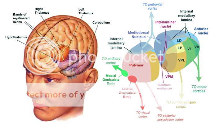
The thalamus is like a switch board. It receives signals from pretty much every part of the brain, sensory, cerebellar, cerebrum, you name it. It also SENDS signals, to pretty much every part as well! This is relevant, as the next door neighbor, the Hypothalamus, as well as Thalamic nuclei, send signals from our actual eyes directly to the Pineal gland, as mentioned earlier by the photo-reception of blue light. Quite an important piece of the puzzle, if you ask me.
Another fact, the Pineal gland happens to sit right at the hind end and wedged inside the two thalamic hemispheres.
The Egyptians seem to have found some significance in this region:

Is it possible that the Pineal, along with the Cerebellum, can help not only coordinate bodily movements, but coordinate movements within the cortex, as well?
Makes you wonder about all that "coincidence" jargon...
Now, mind you, we have to consider that the brain, in it's seemingly infinite complexity, is a highly differentiated device, meaning it is composed of many different macro structures, which they themselves are made up of mostly neurons and other cells which are strongly electrochemical in nature (electromagnetic).
That being said, I'd like to go into the Pineal Gland a wee bit.
Some of you are familiar with the Pineal Gland, and how it is a "third eye" in a sense. The Pineal gland has remnant features that are photo or photon recepting cells within it's body. This is due to fact that the Pineal gland is directly related to Sleep, consciousness, and the circadian cycle.
See, what happens is the Pineal Gland produces Melatonin by default. However, when the wavelengths of optical light, I believe somewhere around 520-540 nm (which is blue light) enter your eyes, those specific photons, of that specific energy, cause the Pineal Gland to shut off Melatonin production. Ever heard of blue blocker lenses? People often will wear these an hour or two before sleep so that they are sleepier.
As for dream time. Well, it's highly possible that the Pineal gland to send out specific triggers, but, if you want to get a better understanding of the matter, you need to look at what the Pineal Gland is connected to, and what those things are directly influenced by.
More on the Pineal's make up:
Pinealocytes: The pinealocytes consist of a cell body with 4–6 processes emerging. They produce and secrete melatonin. The pinealocytes can be stained by special silver impregnation methods. Their cytoplasm is lightly basophilic. With special stains, pinealocytes exhibit lengthy, branched cytoplasmic processes which extend to the connective septa and its blood vessels.
Interstitial cells: Interstitial cells are located between the pinealocytes. They have elongated nuclei and a cytoplasm which is stained darker than that of the pinealocytes.
Perivascular phagocyte: Many capillaries are present in the gland, and perivascular phagocytes are located close to these blood vessels. The perivascular phagocytes are antigen presenting cells.
Pineal neurons: In higher vertebrates neurons are located in the pineal gland. However, these are not present in rodents.
Peptidergic neuron-like cells: In some species, neuronal-like peptidergic cells are present. These cells might have a paracrine regulatory function.
Wha-wha whaaaat? We're talking 4 different types of cells in this puny gland, some of which are neurons! That's one hard working gland.
Now for the Pineal's associations:
If you notice, the Pineal gland (in red) has a red "stalk" that travels down into the very center of the Cerebellum. I'm fairly certain this "Stalk" is a stalk of white matter communicating signals from the Pineal to the Arbor Vitae, which is the central white matter section of the cerebellum. The cerebellum is lightly understood to control muscle coordination, but more recent research has suggested that it sends signals to other parts of the brain, not only the body, which leads me to believe it may just be related to COORDINATION, or TIMING in general.
Furthermore, the simple fact that the pineal gland has a channel going directly into the center of the Cerebellum (which has up to 4 times more processing matter "Gray matter" than the Cerebrum, despite only making up 10% of the brain's volume), combined with the fact that the the cerebellum has, at the center, a seemingly random snake-like portion of gray matter, leads to more questions about the Pineal and it's integration with these structures.
Let's go deeper.
Ever gotten an MRI of your brain? If so, check out the one image that is a side profile, where you can see all of the central structures. I'd post mine, but there's no way I feel comfortable with some certain people seeing that ish...
If you look at it, you'll see what I was talking about above, the Pineal gland attaching to the cerebellum by a stalk. However, you'll also notice that the Pineal gland has two, not one stalk coming off of the front of it as well. This stalk travels through the two Thalami, the two hemispheres of the Thalamus. The region between being known as the cerebro-spinal fluid filled "Third Ventricle".
So now we have the Pineal gland that is directly tied into the center of the white matter inside the cerebellum, and the Pineal also has a stalk that exchanges fluids with the Third ventricle which sits inside the two thalami. In fact:
Unlike much of the rest of the mammalian brain, the pineal gland is not isolated from the body by the blood–brain barrier system;[13] it has profuse blood flow, second only to the kidney.
Curious about what a Thalamic hemisphere is/does?
The thalamus is like a switch board. It receives signals from pretty much every part of the brain, sensory, cerebellar, cerebrum, you name it. It also SENDS signals, to pretty much every part as well! This is relevant, as the next door neighbor, the Hypothalamus, as well as Thalamic nuclei, send signals from our actual eyes directly to the Pineal gland, as mentioned earlier by the photo-reception of blue light. Quite an important piece of the puzzle, if you ask me.
Another fact, the Pineal gland happens to sit right at the hind end and wedged inside the two thalamic hemispheres.
The Egyptians seem to have found some significance in this region:
Is it possible that the Pineal, along with the Cerebellum, can help not only coordinate bodily movements, but coordinate movements within the cortex, as well?
The cerebellum does not initiate movement, but it contributes to coordination, precision, and accurate timing
Makes you wonder about all that "coincidence" jargon...
edit on 19-11-2012 by ProperlyErrant because: (no reason given)
edit on 19-11-2012 by ProperlyErrant because: (no reason
given)
Curious. Interested. Looking for more.
I'll start on ATS, I think.
I'll start on ATS, I think.
Very nice.
The pineal is like the 'third eye' with its sensitive photo-receptive plasma circulation ~
Coordination...coincidences...definitely coordinating coincides now within and between the hemisphere at this little 'eye of ra'!
Thanks
∞LOVE
mayallsoulsbefree∞
The pineal is like the 'third eye' with its sensitive photo-receptive plasma circulation ~
Coordination...coincidences...definitely coordinating coincides now within and between the hemisphere at this little 'eye of ra'!
Thanks
∞LOVE
mayallsoulsbefree∞

reply to post by Druscilla
According to your and "their" quantum level of perception.
"It is easier to stick head in sand, than build sand castle"
-Ancient Chinese Proverb
*yoda snarky voice* mmmhmmhmmhmmhmmmmmm
According to your and "their" quantum level of perception.
"It is easier to stick head in sand, than build sand castle"
-Ancient Chinese Proverb
*yoda snarky voice* mmmhmmhmmhmmhmmmmmm
edit on 20-11-2012 by ProperlyErrant because: (no reason given)
reply to post by ProperlyErrant
Great thread thank you for posting. Photograph and illustrations are very easy to understand. As I am learning about the pineal gland. What have you learned about the effects of sodium fluoride on the pineal gland?
I have read information that suggests that sodium fluoride has a 'hardening' effect on the pineal gland that renders it numb or inactive.
Much Peace...

reply to post by Amanda5
it calcifies in the pineal gland itself.. "blinding" it..
What have you learned about the effects of sodium fluoride on the pineal gland?
it calcifies in the pineal gland itself.. "blinding" it..
Will be very cool if this thread stays up, complete with strassman interview
Commenting to subscribe. Nice OP.
Commenting to subscribe. Nice OP.

Originally posted by Druscilla
reply to post by ProperlyErrant
All I got to say about the Pineal Gland:
Ummmm, Who said anything about supernatural POWERS?????
Where do people get this shiz from ?
I mean, that's quite a leap don't you think?
Someone does a great thread about the Pineal and its obvious connection to some ancient Egyptian culture as well as breaking down some pretty damn good biology of the brain and we don't get three posts deep before someone starts talking about "supernatural POWERS" ?!?!?
Did I miss a post or something?
Who was your comment directed at?
edit on 20-11-2012 by Screwed because: (no reason given)
Amazing post, thanks for shearing your knowledge. I was blown away when I saw the similarities between the eye of ra and the gland. The pineal is a
eye, just as you said is able to detect light. What are your senses? What do they do? They PICK UP and read frequency's with in a certain RANGE.
Their are infinite ranges of frequency's, our 5 senses cannot detect every range. For example some animals cannot see color because their eyes do not
pick up the color's frequency. How wonder how much we are missing because we cannot sense it with our 5 senses. Is the pineal a suppressed 6th
sense? I believe so....
What do you think and more importantly WHY?
What do you think and more importantly WHY?
Originally posted by Druscilla
reply to post by ProperlyErrant
All I got to say about the Pineal Gland:
Did you even read the thread?
Saying
"All I got to say about...." makes it seems as if you did not.
Maybe you will learn something new. DENNY IGNORANCE, open your mind have a look at the information presented.
Posting a home made chart does not prove any thing. Have you looked at any supernatural experiments? And im not talking about those brainwashing tv shows on discovery that are only made to make the supernatural seem unreal. Reveres physiology is one of their most used methods. So have you looked into the supernatural?
edit on 20-11-2012 by Infi8nity because: (no reason given)
Originally posted by Diligence
reply to post by reeferman
Any known methods of de-calcifying it?
Have peek over here ---> My story of de-calcification
On topic: Great thread PE. Very detailed and, although I struggled with the big, confusing words, I feel like i've learnt something tonight. s&f
edit on 20-11-2012 by SilentE because: (no reason given)
new topics
-
Protect the seniors!
Rant: 5 minutes ago -
In another world, it was the apocalypse.
Short Stories: 2 hours ago -
Blinken indicates he would decline any offer to stay on under Harris
Politicians & People: 5 hours ago -
Trump’s N.Y. hush money sentencing delayed until after Nov. election
2024 Elections: 5 hours ago -
is this why they want to go to mars?
Space Exploration: 5 hours ago -
Harris economic plans all seem to involve giving money away
US Political Madness: 6 hours ago -
Israel Defense Forces Shoot American Woman in the Head in Occupied West Bank
Middle East Issues: 7 hours ago -
Secrets of the Name of God & His Epithets (Titles) in the Holy Bible. Life Changing Stuff...
Conspiracies in Religions: 11 hours ago
top topics
-
Trump’s N.Y. hush money sentencing delayed until after Nov. election
2024 Elections: 5 hours ago, 15 flags -
Father of teen suspect in Georgia school shooting charged with 2nd-degree murder
Mainstream News: 17 hours ago, 14 flags -
Harris economic plans all seem to involve giving money away
US Political Madness: 6 hours ago, 10 flags -
Israel Defense Forces Shoot American Woman in the Head in Occupied West Bank
Middle East Issues: 7 hours ago, 8 flags -
is this why they want to go to mars?
Space Exploration: 5 hours ago, 6 flags -
Secrets of the Name of God & His Epithets (Titles) in the Holy Bible. Life Changing Stuff...
Conspiracies in Religions: 11 hours ago, 5 flags -
Blinken indicates he would decline any offer to stay on under Harris
Politicians & People: 5 hours ago, 4 flags -
In another world, it was the apocalypse.
Short Stories: 2 hours ago, 3 flags -
Protect the seniors!
Rant: 5 minutes ago, 0 flags
active topics
-
Are the invader cells being activated? I think so.
Posse Comitatus • 459 • : xuenchen -
KAMALA HARRIS is Having a Hard Time Readying for the 9-10-2024 Debate with Donald Trump.
2024 Elections • 84 • : WeMustCare -
Secrets of the Name of God & His Epithets (Titles) in the Holy Bible. Life Changing Stuff...
Conspiracies in Religions • 12 • : charlest2 -
Protect the seniors!
Rant • 0 • : Bluntone22 -
Democrat VP Candidate Tim Walz Issued Subpoena For $250M COVID-19 Nonprofit ‘Fraud Scheme’
2024 Elections • 17 • : xuenchen -
Due to Trump Leading the Race for President - NY Judge Merchan Will Sentence Him to Prison 9.18.2024
US Political Madness • 57 • : WeMustCare -
is this why they want to go to mars?
Space Exploration • 24 • : tamusan -
This is an anti-free speech right/Nazi/Right Conservatives, a Pro-LGBT community that supports BLM
Rant • 44 • : strongfp -
Blinken indicates he would decline any offer to stay on under Harris
Politicians & People • 20 • : bluesman023 -
Mood Music Part VI
Music • 3581 • : paviabari