It looks like you're using an Ad Blocker.
Please white-list or disable AboveTopSecret.com in your ad-blocking tool.
Thank you.
Some features of ATS will be disabled while you continue to use an ad-blocker.

The Triune Brain and Beyond
page: 17
share:
The Triune Brain and Beyond
Hello ATS readers and contributors. Its been a short while since I have entered this portal, and this will most likely be my last post and visit for a good duration.
Now, onto the subject matter.
Through the last several centuries, there has been a great deal of research into the structures of the brain, as there are notable structures and stratification. As to what the structures do, only in the last several decades have we even begun to figure these notions.
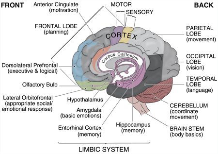
First, I would like to give a brief break down of the “Three” stratified layers of the human brain, as postulated by neuroscientist Paul D. Maclean in his “Triune Brain” theory:
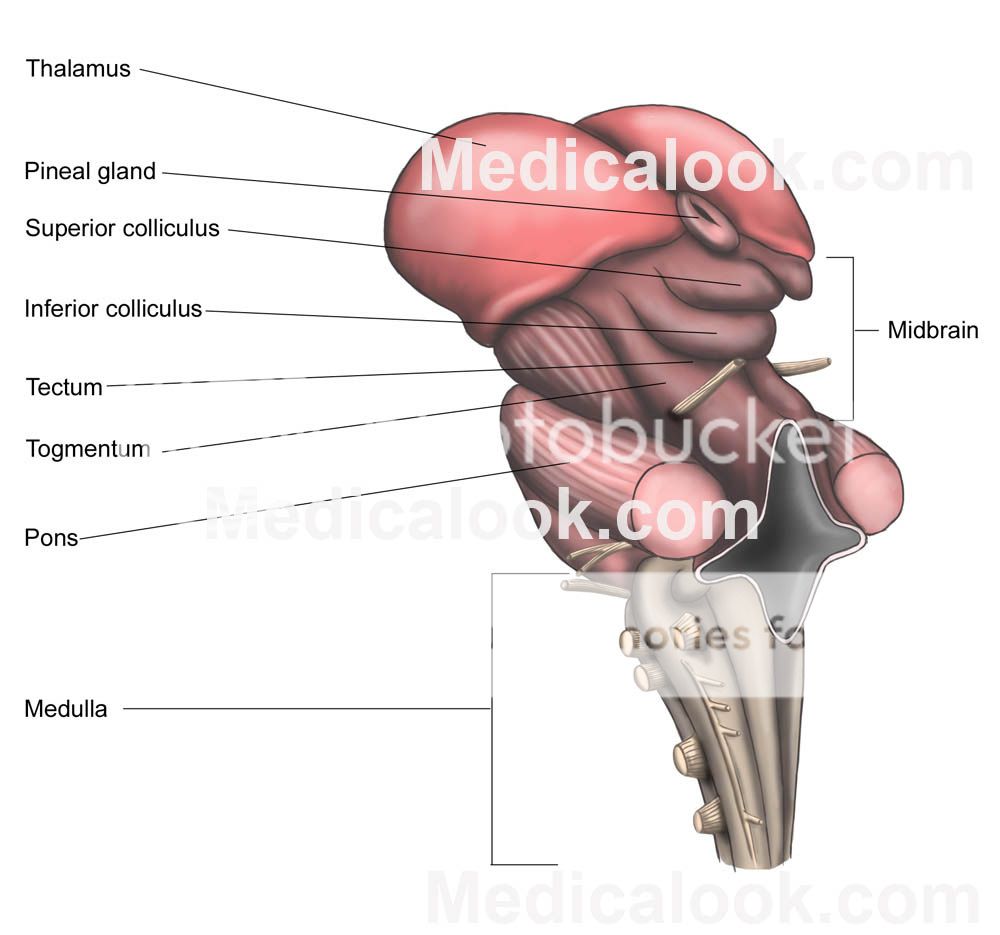
This set of categorizations, of course, leaves out the brain stem, which is comprised of the medulla oblongata (myelencephalon), pons (part of metencephalon), and midbrain (mesencephalon).
The brain stem, is essentially the bridge in which the body (spinal cord) as well as the Cerebellum, are connected, as seen in the above image.
While I will not be going into much of the above mentioned structures, I must add that the Cerebellum, is a fascinating part of the brain that has developed in almost all species, and is most notably responsible for motor control. The cerebellum is tied into, functionally speaking, to the entire body and brain, through its connection to the pons.
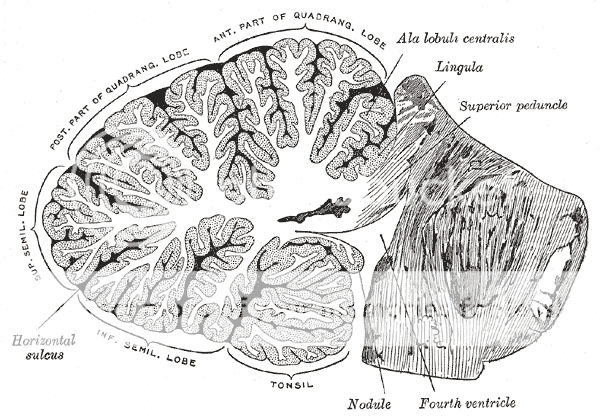
Another fascinating aspect of the Cerebellum is its appearance. Rather than the convoluted, small intestine-looking quality of the neo cortex (The outer most part of the human brain), the Cerebellum instead has parallel grooves, almost resembling muscle fibers, which seems fitting, as the Cerebellum is highly responsible for motor coordination, and function! However, far from being just a muscle, the Cerebellum is incredibly efficient, as it has within it more neurons than the rest of the brain as a whole, yet the Cerebellum only accounts for roughly 10% of the total brain volume. Another incredible feat of this little guy: there are approximately 3.6 times more neurons in the cerebellum than the neocortex! Truly incredible!
...
Now, I know some of you have found the above information enlightening, and good if you have. I will now, however, delve into the more abstract realm.
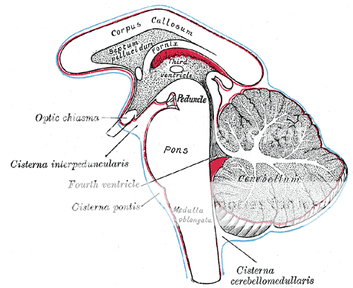
If one were take an exact slice, or rather, one hemisphere of the brain, you will find many of the structures mentioned above, neatly seen in an almost two dimensional format.
Notwithstanding, some argue that the ancient Egyptians already recognized some great importance of the centrally located parts of the brain:

The above image is a hieroglyph of the Eye of Horus/Ra and the internal, central structures of the human brain. Specifically, the comparison is suggesting that the eye of Ra represents these brain structures:
-The Thalamus (central and elliptical red portion)
-The corpus collosum (the thick, white “eye brow”)
-The Fornix (The upper eyelid)
-The brain stem and Cerebellum (The tail trailing to the left and the spiral)
-The Hippocampus (Lower eye lid)
-The Hypothalamus and Optic Chasm (The portion that projects directly downward from the center)
While not exact, and many of you will see no significance due to this lack of exactness, the similarities, as is seen in the similarities of galaxies, solar systems, and atoms, is significant to some of us.
With that said, I would like to delve into one of these structures:
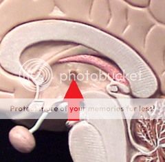
The Thalamus.
While many believe the Pineal Gland to be the center of the brain, and to be this mystical portal to the netherworld (which it may be, as it is related to melatonin, and contains eye-like structures within itself) I find the Thalamus to be particularly fascinating, as it seems to be the center or “eye” of the above mentioned Eye of Ra. So, let’s take a look, shall we?
Hello ATS readers and contributors. Its been a short while since I have entered this portal, and this will most likely be my last post and visit for a good duration.
Now, onto the subject matter.
Through the last several centuries, there has been a great deal of research into the structures of the brain, as there are notable structures and stratification. As to what the structures do, only in the last several decades have we even begun to figure these notions.
First, I would like to give a brief break down of the “Three” stratified layers of the human brain, as postulated by neuroscientist Paul D. Maclean in his “Triune Brain” theory:
Reptilian complex:
The reptilian complex, also known as the R-complex or "reptilian brain" was the name MacLean gave to the basal ganglia, structures derived from the floor of the forebrain during development. The term derives from the fact that comparative neuroanatomists once believed that the forebrains of reptiles and birds were dominated by these structures. MacLean proposed that the reptilian complex was responsible for species typical instinctual behaviors involved in aggression, dominance, territoriality, and ritual displays.
The main components of the basal ganglia are the striatum, the globus pallidus, the substantia nigra, and the subthalamic nucleus. The basal ganglia are a collection of distinct masses of gray matter lying deep in the brain not far from the junction of the thalamus.
The largest component, the striatum, receives input from many brain areas but sends output only to other components of the basal ganglia. The pallidum receives input from the striatum, and sends inhibitory output to a number of motor-related areas. The substantia nigra is the source of the striatal input of the neurotransmitter dopamine, which plays an important role in basal ganglia function. The subthalamic nucleus receives input mainly from the striatum and cerebral cortex, and projects to the globus pallidus
Paleomammalian complex:
The paleomammalian brain consists of the septum, amygdala, hypothalamus, hippocampal complex, and cingulate cortex. MacLean first introduced the term "limbic system" to refer to this set of interconnected brain structures in a paper in 1952. MacLean's recognition of the limbic system as a major functional system in the brain has won wide acceptance among neuroscientists, and is generally regarded as his most important contribution to the field. MacLean maintained that the structures of the limbic system arose early in mammalian evolution (hence "paleomammalian") and were responsible for the motivation and emotion involved in feeding, reproductive behavior, and parental behavior.
Neomammalian complex:
The neomammalian complex consists of the cerebral neocortex, a structure found uniquely in mammals. MacLean regarded its addition as the most recent step in the evolution of the mammilian brain, conferring the ability for language, abstraction, planning, and perception.
This set of categorizations, of course, leaves out the brain stem, which is comprised of the medulla oblongata (myelencephalon), pons (part of metencephalon), and midbrain (mesencephalon).
The brain stem, is essentially the bridge in which the body (spinal cord) as well as the Cerebellum, are connected, as seen in the above image.
While I will not be going into much of the above mentioned structures, I must add that the Cerebellum, is a fascinating part of the brain that has developed in almost all species, and is most notably responsible for motor control. The cerebellum is tied into, functionally speaking, to the entire body and brain, through its connection to the pons.
The unusual surface appearance of the cerebellum conceals the fact that most of its volume is made up of a very tightly folded layer of gray matter, the cerebellar cortex. It has been estimated that, if the human cerebellar cortex were completely unfolded, it would give rise to a layer of neural tissue about 1 meter long and averaging 5 centimeters wide — a total surface area of about 500 square cm, packed within a volume of dimensions 6 cm × 5 cm × 10 cm.[4] Underneath the gray matter of the cortex lies white matter, made up largely of myelinated nerve fibers running to and from the cortex. Embedded within the white matter — which is sometimes called the arbor vitae (Tree of Life) because of its branched, tree-like appearance in cross-section — are four deep cerebellar nuclei, composed of gray matter
Another fascinating aspect of the Cerebellum is its appearance. Rather than the convoluted, small intestine-looking quality of the neo cortex (The outer most part of the human brain), the Cerebellum instead has parallel grooves, almost resembling muscle fibers, which seems fitting, as the Cerebellum is highly responsible for motor coordination, and function! However, far from being just a muscle, the Cerebellum is incredibly efficient, as it has within it more neurons than the rest of the brain as a whole, yet the Cerebellum only accounts for roughly 10% of the total brain volume. Another incredible feat of this little guy: there are approximately 3.6 times more neurons in the cerebellum than the neocortex! Truly incredible!
...
Now, I know some of you have found the above information enlightening, and good if you have. I will now, however, delve into the more abstract realm.
If one were take an exact slice, or rather, one hemisphere of the brain, you will find many of the structures mentioned above, neatly seen in an almost two dimensional format.
Notwithstanding, some argue that the ancient Egyptians already recognized some great importance of the centrally located parts of the brain:
The above image is a hieroglyph of the Eye of Horus/Ra and the internal, central structures of the human brain. Specifically, the comparison is suggesting that the eye of Ra represents these brain structures:
-The Thalamus (central and elliptical red portion)
-The corpus collosum (the thick, white “eye brow”)
-The Fornix (The upper eyelid)
-The brain stem and Cerebellum (The tail trailing to the left and the spiral)
-The Hippocampus (Lower eye lid)
-The Hypothalamus and Optic Chasm (The portion that projects directly downward from the center)
While not exact, and many of you will see no significance due to this lack of exactness, the similarities, as is seen in the similarities of galaxies, solar systems, and atoms, is significant to some of us.
With that said, I would like to delve into one of these structures:
The Thalamus.
While many believe the Pineal Gland to be the center of the brain, and to be this mystical portal to the netherworld (which it may be, as it is related to melatonin, and contains eye-like structures within itself) I find the Thalamus to be particularly fascinating, as it seems to be the center or “eye” of the above mentioned Eye of Ra. So, let’s take a look, shall we?
[The Thalamus’] function includes relaying sensory and motor signals to the cerebral cortex,[2][3] along with the regulation of consciousness, sleep, and alertness. The thalamus has multiple functions. It may be thought of as a kind of switchboard of information. It is generally believed to act as a relay between a variety of subcortical areas and the cerebral cortex. In particular, every sensory system (with the exception of the olfactory system) includes a thalamic nucleus that receives sensory signals and sends them to the associated primary cortical area.
It’s beginning to become clear that this area of the brain is extremely important!
The thalamus also plays an important role in regulating states of sleep and wakefulness.[9] Thalamic nuclei have strong reciprocal connections with the cerebral cortex, forming thalamo-cortico-thalamic circuits that are believed to be involved with consciousness. The thalamus plays a major role in regulating arousal, the level of awareness, and activity. Damage to the thalamus can lead to permanent coma.
Ok, so the Thalamus is EXTREMELY important!
It goes on to say that research in the area of how the Thalamus interacts with some of the midbrain/brain stem structures is almost entirely ignored, including it’s interaction with vestibular and tectal functions, which involves auditory and visual reflexes, as well as balance and spatial orientation, including eye movement and direction - some very important aspects of our functioning as human beings.
The thalamus has been thought of as a "relay" that simply forwards signals to the cerebral cortex. Newer research suggests that thalamic function is more selective
I concur, unequivocally. I often wonder if this section of the brain almost has a “mind” of it’s own, especially considering that it is the most closely connected and oriented portion of the brain to the Pineal Gland! Considering that it has a major interaction with every single sensory system, aside from smell, I’d say it’s pretty damn important. Combined with the fact that the Thalamus is directly and functionally tied into the Hippocampus (which deals with long term, short term memory, and spatial awareness), wow is all I have to say. [As another fascinating fact, did you know that the Pineal Gland is de-activated (in terms of producing Melatonin for sleep) by wavelengths in the blue range of optical light, being 460 to 480 nanometers?]
Kayumov et al. showed that light containing only wavelengths greater than 530 nm does not suppress melatonin in bright-light conditions.[33] Use of blue-blocking goggles the last hours before bedtime has also been advised for people who need to adjust to an earlier bedtime, as melatonin promotes sleepiness.[34]
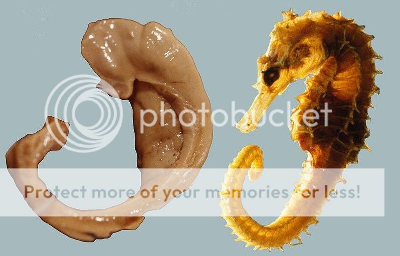
And for yet another image relation, check out the Hippocampus/Fornix complex, as compared to a…Seahorse!
Finally, I’d like to finish up with a tiny, Pineal sized structure in the brain known as the Interthalamic Adhesion. This part of the brain, is a pea sized structure that resides at the center of the two halves of the Thalamus.
In non-human mammals it is a large structure. In humans this mass averages about 1 cm in length in its antero-posterior diameter. It sometimes consists of two parts and occasionally is absent. The interthalamic adhesion is found in 70-80% of humans. It is present more often in females and larger than in males by an average of 53 percent. [1] When absent in development, no noticeable deficit has been observed.
It is theorized that this neuron-laden structure acts almost the corpus collosum does – a channel for connecting the two hemispheres, or in this case, the thalamus halves.
In 1889, a Portuguese anatomist by the name of Macedo examined 215 brains, showing that male humans are approximately twice as likely to lack an interthalamic adhesion as are female humans. He anecdotally attributed the finding to a "prevailing feature of people deprived of [the interthalamic adhesion] is to present in their psychical acts a remarkable precipitation, joined to a certain dysharmony between internal and external feelings."[1] It is noted to be enlarged in patients with Chiari II malformations. [2]
Curious, indeed!
reply to post by Soloro
As for the significance of this “Adhesion”, there is much more research to be done in that area. I did find a bit of research regarding the thickness of the Interthalamic Adhesion In canines:
Whoa. Demented? Seems like a…strong word. To add to that, bigger is certainly not always better. Resonance, or rather, “just right-ness” seems to be the jive.
The brain is truly a remarkable construction of billions of years of cosmic mojo. With that said, here’s a cat with a colorful slinky on its head:

Hope you enjoyed!
As for the significance of this “Adhesion”, there is much more research to be done in that area. I did find a bit of research regarding the thickness of the Interthalamic Adhesion In canines:
The interthalamic adhesion thickness in both T1- and T2-weighted transverse images were measured in all dogs. The interthalamic adhesion thickness in the normal and demented groups was 6.79 +/- 0.70 and 3.82 +/- 0.79 mm, respectively. The interthalamic adhesion thickness in the demented group was significantly smaller […]suggest that interthalamic adhesion thickness may be a good parameter for evaluating brain atrophy in dogs with cognitive dysfunction.
Whoa. Demented? Seems like a…strong word. To add to that, bigger is certainly not always better. Resonance, or rather, “just right-ness” seems to be the jive.
The brain is truly a remarkable construction of billions of years of cosmic mojo. With that said, here’s a cat with a colorful slinky on its head:
Hope you enjoyed!
reply to post by Soloro
Humans have four brains; there is one in the heart. Many heart transplant patients have taken on personality traits and diction patterns, that were those of the donor. Human perception originates with the heart, which then conveys this to the brain that constructs the appropriate sensory impressions.
Humans have four brains; there is one in the heart. Many heart transplant patients have taken on personality traits and diction patterns, that were those of the donor. Human perception originates with the heart, which then conveys this to the brain that constructs the appropriate sensory impressions.
The cat ruined it for me.
Oh, BTW... Star and Flag. I found it interesting.
Oh, BTW... Star and Flag. I found it interesting.
edit on 15-10-2012 by butcherguy because: (no reason given)
reply to post by VoidHawk
I think it's absolutely fascinating how important the Thalamus may actually be, especially regarding it's direct placement with the Pineal. That in itself, almost makes the idea of animals, especially our favorite domestics, seem much more capable and "in tune", rather than disregarding their intelligence, simply because they lack a large neocortex.
I think it's absolutely fascinating how important the Thalamus may actually be, especially regarding it's direct placement with the Pineal. That in itself, almost makes the idea of animals, especially our favorite domestics, seem much more capable and "in tune", rather than disregarding their intelligence, simply because they lack a large neocortex.
new topics
-
Dugin's interview and the West's complete ignorance of Russia
New World Order: 2 hours ago -
Thousands of Anti-Semitic People Protest Israel's government in Tel Aviv
Middle East Issues: 2 hours ago -
Mysterious Spiral 'UFO' Sightings Reported Across US, Europe
Aliens and UFOs: 9 hours ago
top topics
-
The Dark Pyramid of Alaska and the Why Files take on the subject
Whistle Blowers and Leaked Documents: 13 hours ago, 11 flags -
Mysterious Spiral 'UFO' Sightings Reported Across US, Europe
Aliens and UFOs: 9 hours ago, 3 flags -
Thousands of Anti-Semitic People Protest Israel's government in Tel Aviv
Middle East Issues: 2 hours ago, 3 flags -
Dugin's interview and the West's complete ignorance of Russia
New World Order: 2 hours ago, 3 flags
active topics
-
For Votes - President BIDEN Opens ObamaCare Health Ins to Illegal Aliens Eff Nov 1st 2024.
2024 Elections • 26 • : WeMustCare -
Canada caught red-handed manipulating live weather data and make it warmer
Fragile Earth • 45 • : leongrad -
Poll - Catholic Support Swings to Trump By Significant Margin
2024 Elections • 70 • : andy06shake -
Really Unexplained
Paranormal Studies • 86 • : Beesnestbomber -
Israeli strikes on southern Gaza city of Rafah kill 22, mostly children, as US advances aid package
Middle East Issues • 138 • : Dalamax -
The Vaccine Injured
Medical Issues & Conspiracies • 21 • : PrivateAngel -
The Dark Pyramid of Alaska and the Why Files take on the subject
Whistle Blowers and Leaked Documents • 5 • : sine.nomine -
Mysterious Spiral 'UFO' Sightings Reported Across US, Europe
Aliens and UFOs • 11 • : Zaphod58 -
Thousands of Anti-Semitic People Protest Israel's government in Tel Aviv
Middle East Issues • 11 • : gortex -
Zionists of ATS assemble
Political Issues • 168 • : CriticalStinker
7